Mayo Clinic (May 11, 2023) – In the U.S., HPV is linked to about 70% of throat and mouth cancers. And more than 70% of those cancers are diagnosed in men, according to the Centers for Disease Control and Prevention.
Treatment for throat and mouth cancers, also referred to as oropharyngeal or head and neck cancers, will depend on location and stage of the cancer as well as other factors. Dr. Phillip Pirgousis, a Mayo Clinic head and neck surgeon, says patients have safer, less invasive surgical treatments available to them thanks to ongoing innovation.
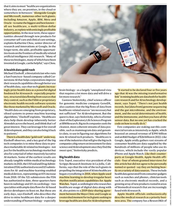
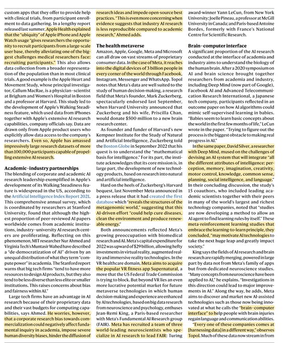
Scripps Research (April 11, 2023) – From smartwatches and fitness bands to glucose monitors and in-home ultrasounds, the proliferation of digital devices is igniting a revolution in healthcare and medical research.
Patients can now collect thousands of data points about themselves and share that information with their healthcare providers. At the Scripps Research Translational Institute, researchers are taking advantage of new technology to study disease in novel ways.
Their projects include a platform for early detection of disease outbreaks, a sleep quality study, and even a way to predict and individual’s risk of certain disease based on their genetics. In this video, hear directly from the team about this exciting new frontier.
Northwestern Medicine (March 21, 2023) – Parambir Dulai, MD, is studying the use of hyperbaric oxygen therapy to treat severe ulcerative colitis. The treatment delivers oxygen into the tissues to promote healing.
What Is Ulcerative Colitis?
Ulcerative colitis is an inflammatory bowel disorder that affects the large intestine and rectum, causing bleeding. This chronic condition impacts more than 900,000 Americans. It can begin at any age, but it often starts in young adulthood. Older men are more likely to be diagnosed than older women.
There is no cure for ulcerative colitis, but with treatment, the majority of patients can be symptom-free for long periods of time.
After a digital twin of a heart is created, researchers can go a step further and use 3D printing to create a physical version of a heart. This is then used to practice surgical techniques and test solutions such as new heart valves or drugs without ever touching an actual body.
March 2, 2023: Following National Heart Health Month in February, TCS futurists took a look at how a digital twin of the heart can save more lives – human and animal – in the future. From boosting athletic performance to developing predictive medicine, new advances in technology will help keep hearts healthier than ever.
TCS is on the leading edge of “Digital BioTwin” research, modeling human organs digitally to find new ways for researchers and doctors to test experimental drugs and surgical techniques without risk. With heart disease the leading cause of death in the U.S., it is more important than ever to innovate techniques to keep hearts healthy.
Using information from a MRI of someone’s heart, TCS can create a fully modeled human heart in cyberspace. By applying various historical and speculative data sets, doctors can see the impact of different conditions and situations such as beginning a long-term exercise program or quitting smoking. This approach to predictive medicine demonstrates the real impact of health choices to patients.
Yale Medicine (February 11, 2023) – Robotic bronchoscopy, also known as robotic-assisted bronchoscopy, is a recent advancement in bronchoscopy, the procedure used to biopsy lung nodules to detect the presence of lung cancer and other lung diseases. Lung cancer is the third most common type of cancer and the leading cause of cancer-related death in the United States.
Early diagnosis and treatment often lead to better outcomes. A diagnosis of lung cancer often begins when a chest X-ray or computed tomography (CT) scan shows a nodule—an area of abnormal tissue—in the lungs. If the nodule is suspicious or grows over time, doctors will perform a biopsy to collect a tissue sample that can be tested for the presence of cancer cells. Bronchoscopy is a widely used procedure for biopsying lung nodules.
In traditional bronchoscopy, a doctor manually guides a thin tube, called a bronchoscope, into the patient’s mouth or nose, down the throat, past the vocal cords and windpipe, and into the passageways of the lungs. The bronchoscope is equipped with a light, camera, and biopsy tools that allow doctors to visually examine and biopsy nodules. But robotic bronchoscopy is different. Like traditional bronchoscopy, it’s a minimally invasive procedure that allows doctors to biopsy nodules in the lungs.
The difference is that in robotic bronchoscopy, the doctor uses a controller at a console to operate a robotic arm. The robotic arm guides a catheter—a thin, flexible, and maneuverable tube equipped with a camera, light, and shape-sensing technology—through the patient’s airways. The robotic arm’s precise movements enable doctors to accurately direct the catheter around tight turns in the airways and into the hard-to-reach areas of the lungs. This means doctors can examine and biopsy suspicious nodules—and potentially detect cancer—in parts of the lungs that may be inaccessible with traditional bronchoscopy.
What’s more, the procedure is safe—serious complications are rare—and recovery is usually quick. “As part of the comprehensive Thoracic Oncology Program, we are now able to offer patients the option of robotic bronchoscopy,” says Yale Medicine interventional pulmonologist Christopher Morton, MD. “This technology will allow us to biopsy lung nodules and masses with improved accuracy and fewer side effects, in addition to lymph node biopsies that we already do. This will get patients diagnosed and referred to the appropriate treating physician quicker.”
“We found that the information we could get from PSMA scanning in patients with newly-diagnosed prostate cancer before surgery was at least as reliable and useful as other information from biopsy, PSA levels, or clinical exam for predicting how patients would do after surgery or other treatment,” says Farshad Moradi, a radiologist at Stanford who co-authored the study.
In December, scientists at Stanford University reported promising findings with a new technology that lights up prostate tumors on specialized imaging scans. The approach relies on a minimally-radioactive tracer that travels the body hunting for cancer cells.
Called 68Ga-PSMA-11, and delivered intravenously, the tracer binds exclusively with a protein called prostate- specific membrane antigen (PSMA). Prostate cancer cells contain far more of this protein on their surfaces than normal prostate cells do. Tumors flagged by 68Ga-PSMA-11 show up on an imaging scan like lit matches in a dark room. Doctors are already using PSMA scans to diagnose early metastatic cancer, and the tracer can also be used to ferry drugs directly into malignant tumors.
Recently, low-field MRI scanners have become available that are portable, are cryogen-free, are easy to use, provide rapid patient loading and unloading, have minimal power requirements, and have relatively low purchase prices and maintenance costs. For some indications, including ischemic stroke, these MRI scanners are a welcomed addition to the clinical armamentarium, as they have the potential to improve some aspects of clinical care over the current standard of care.
For one, they offer rapid “point-of-care” imaging diagnosis. Owing to their reduced cost and portability, these scanners could be deployed in a myriad of new settings, such as at-large public gatherings (e.g., sporting events or rock concerts), rural health care centers, emergency rooms, and assisted living facilities. Future innovations in motion correction, noise remediation, and image data upload capabilities suggest the eventual use of these scanners in ambulances or even on the battlefield.
Shoulder replacement is a major surgery where all or part of the shoulder joint is replaced. In the past, it would take weeks for the patient to gain mobility, but new technology is changing all that.
Laura Britt has degenerative joint disease and as a result has had several surgeries, including shoulder replacements.
“The data we analyzed suggested a nearly threefold reduction in revision surgery in patients who received bone marrow aspirate concentrate, compared to those who did not,” says Bradley Schoch, M.D., an orthopedic surgeon and principal investigator. “This procedure is growing in use throughout the practice of orthopedic surgery and commonly added as a surgical adjunct to rotator cuff tears.”
Mayo Clinic researchers analyzed the largest set of data available to determine if adding bone marrow aspirate concentrate to repaired tissue after standard rotator cuff surgery would improve outcomes for patients. Bone marrow aspirate is fluid taken from a patient’s bone marrow that contains concentrated growth factors, stem cells and other specialized cells that may regenerate tissue and cartilage.
The analysis identified 760 patients who had a regenerative intervention added to augment rotator cuff repair surgery. Those patients were compared to 3,888 patients who did not have any biologic intervention at the time of surgery. The data indicated that 114 patients who opted for bone marrow aspirate concentrate at the time of surgery were less likely to need a second surgery.