Tag Archives: Pulmonary Circulation

BLOOD VESSELS: HUMAN “PULMONARY CIRCULATION”

Humans have a high energy requirement. Like a sports car we need to be turbosupercharged. We need an entirely separate Pulmonary circulation to handle our great oxygen demand.

Fish can get by on a single heart and circulation. They are “cold-blooded” and have no elevation of temperature above that in the environment. The water buoys them up, and they don’t need to constantly fight gravity.

Birds, and by extension, therapod dinosaurs, need more efficiency, and have a separate pulmonary circulation, just like we do. They share with us a DOUBLE CIRCULATION, a 4-chambered heart, with 2 entirely separate circuits.

In my residency, I saw a lot of congenital heart disease. In the process of development, the very early human embryo has a single circulation, just like “early” vertebrates, like fish.

In the process of development, the Systemic and Pulmonary circulations divide the previously unitary system into 2 separate systems, by a continuous spiral of partitions, or “septae”.

Ontogeny recapitulates Phylogeny: Development recaps Evolution. If this process of separation fails to happen in a given child, Congenital Heart Disease is the result:

  • IASD. Interatrial Septal Defect is failure to separate the Atria, the upper chambers of the heart;
  • IVSD, Interventricular Septal Defect, is failure to separate the Ventricles, the lower chambers of the heart;
  • AV Communis is both of the above, plus failure of Atria and Ventricles, the upper and lower chambers, to separate, giving one big inefficient chamber.

When you listen to the hearts of these children, there are prominent murmurs, or noises, which betray the presence of turbulence and inefficiency, the very thing that evolution “tried” to prevent.

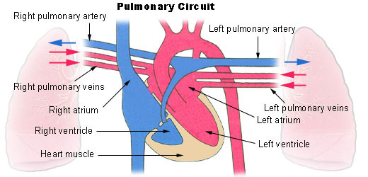
In the normal Human Heart, the blood returns from it’s systemic circuit through the capillaries, depleted of oxygen, into the vena cava. It passes to the Right Atrium, through the tricuspid valves, to the right ventricle.

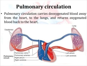
With the contraction of the heart, the blood goes through the pulmonary artery, into the pulmonary capillaries, into close contact with air-containing alveoli. The oxygen passes through the alveolar membrane into the capillary blood, which becomes red. The oxygenated blood then passes into the pulmonary veins and on into the left atrium, and the systemic circulation.

It is interesting, and essential that the systemic arteries contain red, oxygenated blood, and the Pulmonary artery contains blue, oxygen-depleted blood. The “tired” blood, returning from the body must be “pepped up” by passing through the pulmonary circuit, picking up oxygen in the process.

Similarly a clot, originating in a quiet vein, perhaps a dilated, or varicose vein, is pumped into the Pulmonary circuit, where it lodges in the tiny capillaries and produces a PULMONARY EMBOLISM.

The embolus clogs the pulmonary circuit, increasing resistance, raises the normally-low pulmonary artery pressure and produces PULMONARY HYPERTENSION, placing more load and strain on the Right Ventricle.

Pulmonary Hypertension is also caused by a variety of Lung, heart, inherited and kidney diseases, as well as by certain drugs, high altitude, and Obstructive Sleep Apnea.

Please check the Mayo Clinic discussion that follows.

–Dr. C

Pulmonary Hypertension Article

BLOOD VESSELS: THE “SYSTEMIC ARTERIAL SYSTEM”

BLOOD VESSELS; it is hard to overestimate their importance. They are literally our lifelines, delivering the oxygen and nutrition necessary for life. We are as old as our blood vessels.

I will divide blood vessels into 4 components with rather separate domains: The Systemic Arterial system, the Pulmonary circulation, the Venous system, and the Lymphatic system, and will discuss these separately.

SYSTEMIC ARTERIAL SYSTEM

The Boy Scouts taught me the pressure points; The radial, at the thumb-side of the wrist, the brachial, on the inside of the upper arm, and the inguinal in the groin area. Pressure on these sites will stop arterial bleeding distally.

You should be able to locate the radial artery pulse, and begin to appreciate its strength and regularity. Strength in case you encounter a person who isn’t moving, and regularity for yourself; many older people develop an irregularity called Atrial Fibrillation, and you might be the first to discover it..

ANEURISMS are swelling of the arteries, and the swelling may thin the arterial wall so that it can burst. A Cerebral aneurysm can burst and cause a stroke-like problem. If an aortic aneurism bursts, the internal blood loss can be fatal.

RAYNAUD’S Phenomenon is fairly common, and consists of an over-reaction to cold, where arteries of the hands constrict, and the fingers get white and cold. Burger’s disease involves small arteries, and often is associated with Raynaud’s. The arteries carry the blood distally (away from the heart), continuing to divide into ever smaller arterioles which terminate in capillaries, which branch out in such an arborization as to supply all cells except cartilage and parts of the eye.

HYPERTENSION develops when the arterioles, under hormonal or neural influence, constrict, increasing the resistance to blood flow, and so the pressure. Increase in sodium retention and therefore the blood volume can also increase pressure.

ATHEROSCLEROSIS is the common disease of western life style. Excessive calories and sedentary life style combined with genetic defects in fatty metabolism produce cholesterol plaques which narrow and stiffen the arteries, often leading to BLOCKAGE of blood flow. Blockage of flow to the HEART, BRAIN, KIDNEYS, BOWEL, or EXTREMITIES each produce their separate disorders of Myocardial Infarction, Stroke, Renal artery Disease. Intestinal ischemic syndrome, and Claudication.

Atherosclerosis

These disorders will each be separately discussed. I have always thought of vascular disease as a special class of CAUSATIVE MECHANISMS when trying to develop a DIFFERENTIAL DIAGNOSIS of a patient’s problems. Blockage to an area results in PAIN or LOSS OF FUNCTION.

Stroke is usually painless with blockage, since the brain has no pain sensors. Blockage of the renal artery often causes complex difficulties including Hypertension, because the kidney is an endocrine organ in addition to its excretory function.

A good Friend and patient showed what careful self-care can accomplish. It all started with a myocardial infarction, the first sign of his blood vessel disease. He had a complication in his workup, and had to have emergency bypass surgery. There had been damage to the heart muscle, with a large reduction in his EJECTION FRACTION.

His cardiologist gave him at most 5 years to live. That was 25 years ago, before the development of the statin drugs. He was given a draconian low cholesterol diet, which he followed exactly. One one visit to the cardiologist, he inquired whether he could have other areas of arterial blockage. His doctor then listened to his neck and discovered a bruit (noise) in the carotid artery, after which he had a Carotid endarterectomy.

In an orthopedist office for back pain, the orthopedist left the room, and my friend noticed in the CT scan report mention of cysts in the kidneys.

The Orthopedist cared mainly about his bones, and had overlooked the “incidental finding”. His brother had died of mesenteric artery blockage from atherosclerosis, he had stomach symptoms, and sure enough he also had arterial blockage to the intestines.

Bottom line: it pays to be an ACTIVE PARTICIPANT in our medical treatment, and even though we all have genetic determinants, we can make our health BETTER with attention to our health, especially SLEEP, DIET and EXERCISE.

–DR. C