
At one time, replacement parts for the eyes must have seemed unimaginable. Nowadays, if the inner lens of the eye becomes clouded by a cataract, a routine surgery to swap it out with a new artificial lens restores vision.
But what happens if the outer lens of the eye (the cornea) becomes damaged or diseased? You can have that replaced, too. “It’s not as common as cataract surgery, but many people get corneal diseases after age 50 and may need a corneal transplant,” says Dr. Nandini Venkateswaran, a corneal and cataract surgeon at Harvard-affiliated Massachusetts Eye and Ear.
More than 49,000 corneal transplants occurred in 2021 in the US, according to the Eye Bank Association of America.
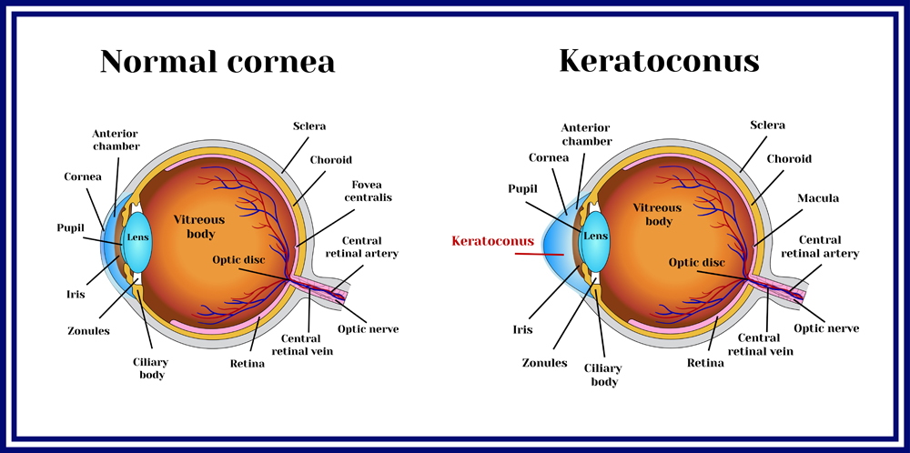
What is the cornea?
The cornea is a dome of clear tissue at the front of each eye, covering the iris and pupil, that acts as a windshield that protects the delicate eye apparatus behind it, and focuses light onto the retina, which sends signals that the brain turns into images (your vision).
You need this combo of windshield and camera lens to focus and see clearly. But many things can go wrong within the five layers of tissue that make up the cornea. That can make it hard to see and rob you of the ability to read, drive, work, and get through other activities in your day.
How does damage to the cornea occur?
It may stem from a number of causes:
- Injuries, such as a fall. “Falls are a big reason for people to come in with acute eye trauma. The cornea can be damaged easily if something pokes it,” Dr. Venkateswaran says.
- Previous eye surgeries. “Especially for adults who’ve had several eye surgeries — such as cataract and glaucoma surgeries — the inner layers of the cornea can become damaged and weakened with age,” she adds.
- Illness. Problems like severe corneal infections, or genetic conditions such as Fuchs’ endothelial dystrophy, can cause vision loss.
What are the options for treating corneal damage?
Cornea treatment depends on the type of problem you have and the extent of the damage. “It’s a stepwise approach. Sometimes wearing a specialty contact lens or using medications can decrease swelling or scarring in the cornea,” Dr. Venkateswaran says.