Category Archives: Infographics

Infographic: Diagnosis & Treatment Of Psoriasis

Psoriasis is a skin disease that causes a rash with itchy, scaly patches, most commonly on the knees, elbows, trunk and scalp.

Psoriasis is a common, long-term (chronic) disease with no cure. It can be painful, interfere with sleep and make it hard to concentrate. The condition tends to go through cycles, flaring for a few weeks or months, then subsiding for a while. Common triggers in people with a genetic predisposition to psoriasis include infections, cuts or burns, and certain medications.

Treatments are available to help you manage symptoms. And you can try lifestyle habits and coping strategies to help you live better with psoriasis.

Website

COMMENTS ON ‘PSORATIC ARTHRITIS’:

My practice was restricted to allergy, but I saw many patients with psoriasis. The red scaly patches made them think they had allergic dermatitis, eczema. Psoriasis on the arm is usually located on the elbow, and atopic dermatitis on the opposite side, in the flexural area. Thick, pitted fingernails are also common in psoriasis. It’s combination with arthritis is worrisome.

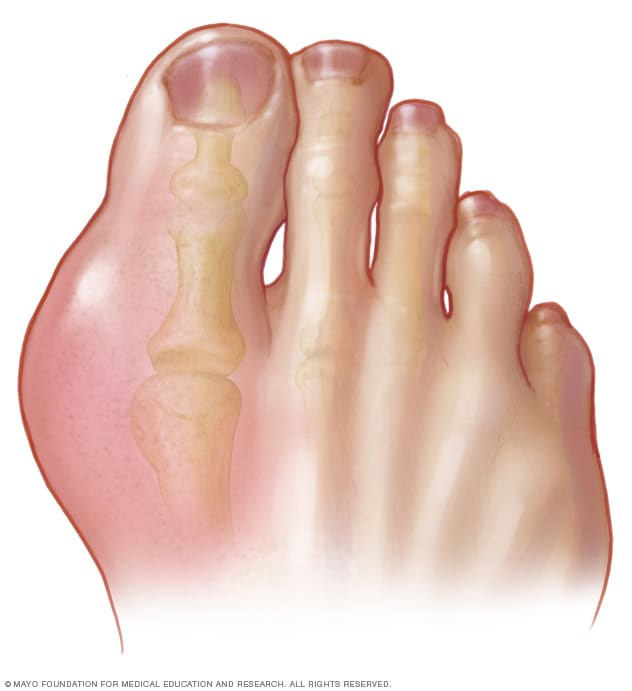
Psoriasis will usually develop first, and the psoriatic arthritis will follow years later, but 10% of the time the arthritis Is the first problem. This form of arthritis can be very painful, and cause deformities. It is often worse than rheumatoid arthritis, although does not affect as many joints, and is often asymmetrical. It inflames the area where tendons attach to the bone, which is one of the reasons that it can be more painful than rheumatoid arthritis.

Psoriasis is an autoimmune problem and can involve practically any organ in the body.

It is often associated with metabolic syndrome and diabetes.

Psoriatic arthritis does not have the rheumatoid serum markers that can help diagnose rheumatoid arthritis, and unless psoriasis is also present on the skin, it can be hard to diagnose.

Symptomatic treatment with NSAIDs, physical therapy, phototherapy and topical treatments can be helpful, but very expensive biologics are sometimes needed to help out methotrexate and other first line DMARDs (Disease modifying antirheumatic drugs).

This condition can be  progressive.  If you develop scaly red patches on your skin, be sure to check with the doctor about the possibility of psoriasis.

—Dr. C.

Bacteria: The Risks Of Pseudomonas Aeruginosa

Bacteria have long been classified according to how they are stained by the chemical dye called the Gram stain. Pseudomonas aeruginosa is Gram-negative, compared to the Staph aureus which is gram-positive.

Being Gram negative, Pseudomonas has an extra membrane, the outer membrane, and a shell of a nasty material called Lipo polysaccharide. These extra structures act as a barrier to entrance of antibiotics. In addition, the Pseudomonas has many more genes than the average bacterium and uses these genes for adaptation. For instance, it is good at forming a raft of mutually supporting cells called a biofilm, which enables it to become particularly entrenched. In a condition of the lung called cystic fibrosis, this biofilm contains specialized cells, one of which is called the Persister cell. When Pseudomonas senses that a critical number of bacteria is present, called quorum sensing, the persister cell multiplies to become around 10% of the bacterial population, and slows down their metabolism massively, becoming a sort of “zombie cell”. These cells are very hard to kill and persist through an antibiotic treatment that kills other Pseudomonas cells, only to rev up their metabolism and become active again once treatment is withdrawn.

In addition, Pseudomonas has all of the other resistant talents mentioned in the previous article on Staphylococcus aureus, such  as plasmid acceptance, ability to destroy penicillin, efflux pumps, and rerouting of metabolism.

In trying to control Pseudomonas, techniques other than antibiotics are being tried out of desperation, including interference with a quorum sensing, use of bacteriophages and chemical  elements such as Gallium which masquerades as the iron this bacterium requires.

Pseudomonas is not as actively pathogenic as Staphylococcus aureus, but it has made a great niche for itself in the respiratory tract, especially in people with a compromised immune system, or pulmonary abnormality such as cystic fibrosis. Ventilation tubes and other hospital equipment can become contaminated and spread the infection, unless thoroughly cleansed of Pseudomonas; it is very sensitive to acidic solutions, and those who need nasal CPAP for sleep apnea may recall that they have to rinse their equipment in vinegar, or acetic acid. Swimmers can get an external ear infection with this organism, and the drops for swimmers ear often contains acetic acid.

The next rogue to consider is a frightful yeast called Candida auris.

—Dr. C.

Infographic: Acute Heart Failure (Nature Reviews)

Acute heart failure (AHF) is a syndrome defined as the new onset (de novo heart failure (HF)) or worsening (acutely decompensated heart failure (ADHF)) of symptoms and signs of HF, mostly related to systemic congestion. In the presence of an underlying structural or functional cardiac dysfunction (whether chronic in ADHF or undiagnosed in de novo HF), one or more precipitating factors can induce AHF, although sometimes de novo HF can result directly from the onset of a new cardiac dysfunction, most frequently an acute coronary syndrome.

Despite leading to similar clinical presentations, the underlying cardiac disease and precipitating factors may vary greatly and, therefore, the pathophysiology of AHF is highly heterogeneous. Left ventricular diastolic or systolic dysfunction results in increased preload and afterload, which in turn lead to pulmonary congestion. Fluid retention and redistribution result in systemic congestion, eventually causing organ dysfunction due to hypoperfusion. Current treatment of AHF is mostly symptomatic, centred on decongestive drugs, at best tailored according to the initial haemodynamic status with little regard to the underlying pathophysiological particularities.

As a consequence, AHF is still associated with high mortality and hospital readmission rates. There is an unmet need for increased individualization of in-hospital management, including treatments targeting the causative factors, and continuation of treatment after hospital discharge to improve long-term outcomes.

COMMENTARY:

High frequency sound (ultrasound) bounces off of tissues, like an echo,  and allows an electronic look at the heart. Doppler echocardiography is the doctors method of choice for evaluating a heart failure. One of the most important numbers determined by this method is the EJECTION FRACTION, which is a measure of heart efficiency. If the ejection fraction is low, let’s say below 45%, the heart is pumping out only 45% of its volume with each stroke, which means it must work harder to produce the same amount of circulation. The normal is about 60%.

This is the basis of SYSTOLIC Heart failure.

The test can also tell about blood coming into the heart; the early part of the blood entering is usually 80% of the total. If it drops, let’s say below 50%, it means the heart is stiff and resists blood coming in, which is the basis of DIASTOLIC heart failure.

FACES Is an acronym-mnemonic for the symptoms of heart failure. F is for FATIGUE.

A is for ACTIVITY LIMITATION. C is for CONGESTION in the lungs.  E is for EDEMA, or swelling, usually of the ankles and legs. S is for SHORTNESS of BREATH.

If you want to remember a bit about heart failure, think about echoes and faces. If your Doctor orders an ultrasound with Doppler, be sure to ask about your ejection fraction and percent of blood that enters early, before the “atrial kick”. Being informed is always a good thing.

To be honest, I have never encountered a person who was given a Doppler echocardiogram and could tell me what his ejection fraction was, but I am eternally hopeful.

—Dr. C.

Arthritic Conditions: The Signs & Symptoms Of Gout

#Gout is a common & complex form of arthritis that can affect anyone. It’s characterized by sudden, severe attacks of pain, swelling, redness and tenderness in joints, most often in the big toe.

An attack of gout can occur suddenly, often waking you up in the middle of the night with the sensation that your big toe is on fire. The affected joint is hot, swollen and so tender that even the weight of the bedsheet on it may seem intolerable.

Gout symptoms may come and go, but there are ways to manage symptoms and prevent flares.

Symptoms

Gout in the big toe

The signs and symptoms of gout almost always occur suddenly, and often at night. They include:

  • Intense joint pain. Gout usually affects the big toe, but it can occur in any joint. Other commonly affected joints include the ankles, knees, elbows, wrists and fingers. The pain is likely to be most severe within the first four to 12 hours after it begins.
  • Lingering discomfort. After the most severe pain subsides, some joint discomfort may last from a few days to a few weeks. Later attacks are likely to last longer and affect more joints.
  • Inflammation and redness. The affected joint or joints become swollen, tender, warm and red.
  • Limited range of motion. As gout progresses, you may not be able to move your joints normally.

Read about the ways to manage symptoms & prevent flares. https://mayocl.in/3P29WFA

COVID-19: HEART DISEASE RISKS RISE AFTER INFECTION

In December 2020, a week before cardiologist Stuart Katz was scheduled to receive his first COVID-19 vaccine, he came down with a fever. He spent the next two weeks wracked with a cough, body aches and chills. After months of helping others to weather the pandemic, Katz, who works at New York University, was having his own first-hand experience of COVID-19.

On Christmas Day, Katz’s acute illness finally subsided. But many symptoms lingered, including some related to the organ he’s built his career around: the heart. Walking up two flights of stairs would leave him breathless, with his heart racing at 120 beats per minute. Over the next several months, he began to feel better, and he’s now back to his normal routine of walking and cycling. But reports about COVID-19’s effects on the cardiovascular system have made him concerned about his long-term health. “I say to myself, ‘Well, is it really over?’” Katz says.

In one study1 this year, researchers used records from the US Department of Veterans Affairs (VA) to estimate how often COVID-19 leads to cardiovascular problems. They found that people who had had the disease faced substantially increased risks for 20 cardiovascular conditions — including potentially catastrophic problems such as heart attacks and strokes — in the year after infection with the coronavirus SARS-CoV-2. Researchers say that these complications can happen even in people who seem to have completely recovered from a mild infection.

Some smaller studies have mirrored these findings, but others find lower rates of complications. With millions or perhaps even billions of people having been infected with SARS-CoV-2, clinicians are wondering whether the pandemic will be followed by a cardiovascular aftershock. Meanwhile, researchers are trying to understand who is most at risk of these heart-related problems, how long the risk persists and what causes these symptoms.

COMMENTARY:

 The heart and Covid are connected from a variety of angles.

Obese people with high blood fats, diabetes, the metabolic syndrome tend to have atherosclerosis and heart problems, making them more susceptible to severe Covid and long Covid. Covid loves to involve the lining of blood vessels and the heart, the endothelium, where the number of ACE receptors are high.

The respiratory tract and lung are a particular target for Covid, and reduced oxygen from lung involvement can compromise the hard-working heart.

 Heart cells, cardio myocytes, can be directly infected with the virus. Even Covid vaccines can rarely produce myocarditis, raising the possibility that there is some antigenic similarity between the virus and heart cells, similar to the beta hemolytic streptococcus and the heart which sets up rheumatic fever. 

If this similarity is real, the tendency of Covid to compromise the immune system and produce a cytokine storm in severe cases could therefore specifically involve the heart.

The nature article indicates several different varieties of heart problems and is a recommended read. From my personal standpoint, arrhythmias were mentioned, and I already have trouble with a couple of different types, AF and NSVT.

To make definite statements about the likelihood of heart involvement in Covid is problematic. The patients reported on were infected with an earlier strain of Covid, and the present one, BA.5, seems to be milder, and  may not be as hard on the heart as previous strains. Many more people are now immunized, and the most susceptible patients may have passed away. There are medications to take, such as remdesivir, and even select immune globulins, such as an immuno-suppressed friend of mine was given when he contracted Covid recently.

The bottom line for me is that I am 90 years old and have no desire to let Covid have a crack at me, so I avoid big gatherings, and wear a mask whenever I am exposed.

—Dr. C.

Read more

Diagnosis: Hypertensive Eye Disease (Infographic)

Hypertensive eye disease is diagnosed on the basis of a clinical history of #Hypertension and the presence of relevant pathological changes in the eye that are observed via fundoscopy or imaging. https://go.nature.com/3t0PahO

Commentary:

Control of hypertension is one of the triumphs of modern medicine, and there are many effective medications. One of the main problems that I hear about has  to do with taking too much of the medication, and having dizziness, especially on standing, which could cause a fall and injuries.

Abnormalities of the eye with hypertension are common. the doctor can look directly at the bare blood vessels as they course along the back of the eye. A hypertensive artery passing over a vein in the eye compresses it, producing a “nick” that doctors look for. other findings are shown in the info graphic.

These findings help doctors make a diagnosis, but only the rare complications of hypertensive optic atrophy and Choroidopathy actually endanger the vision.

The main organ that seems to suffer the most from hypertension is the heart, which has to work against a heavy load to pump the blood effectively at high pressure. This thickens the walls of the heart, especially the left side, making it less effective. Enlargement of the heart, heart attacks, and heart failure are not uncommon.

The brain is at risk with hypertensive arterial disease, and strokes can be a problem.

Kidney failure is also a real worry.

When you see your doctor for a blood pressure reading, make sure to take off coats and long sleeves. so that the bare arm can be tested. The left arm In right-handed people is preferable, because it has less musculature to shield the artery from compression by the blood pressure cuff.

Be sure you take your medication, if prescribed. The main emergency room visits occasioned by high blood pressure, such as 180/110, is when the patient skips the medication.

—Dr. C.

Dr. C’s Journal: MRSA- Methicillin Resistant Staphylococcus Aureus

Staphylococcus aureus is a highly successful – eg.BAD – disease producing bacterium, or “pathogen”. From the standpoint of the bacterium, it didn’t know it was so bad, and was merely producing factors that break down its surroundings to produce food for multiplication. Unfortunately, the proteins-ENZYMES- produced by the staphylococcus, In its effort to survive, can cause anything from diarrhea, abscesses, high fevers, shock, and kidney failure to death.

The staphylococcus was very successful for thousands of years, until slowed down by penicillin.

Penicillin attached itself to a critical part of the staphylococcal cell wall, which stopped the bacterium from functioning. The staphylococcus retaliated by producing an enzyme, penicillinase, that destroys penicillin. Humans responded by producing methicillin, which resisted destruction by the penicillinase. The Staph responded again by a genetic change in the target of penicillin, PBP, so that the Methicillin wouldn’t attach, hence the term methicillin resistant staphylococcus aureus, MRSA.

Interestingly, the MRSA can send little packets of genes called “plasmids” to other bacteria, even other species, which take in the packets and allow them to resist methecillin also. This is called horizontal transfer of resistance.

The staphylococcus aureus has many other ways of protecting itself, and is a good example of the various ways a bacterium can deal with antibiotics.

It can prevent the antibiotic from gaining entrance, or pump it out of the cell faster. It can destroy the antibiotic, as with penicillinase. It can modify the bacterial target of the antibiotic, as in the PBP that protects from penicillin. It can also develop an alternative metabolic pathway or structure to bypass the effect of the antibiotic.

THIS IS WAR, similar to the human kind. The offense develops the sword. The defense develops the shield. The offense develops mounted archery. The defense develops the castle and so on.

We’re not so very different from the bacteria, and in fact every single cell of our bodies has descended from an ancestral  bacterial cell that engulfed another bacterium, which became the mitochondrion, a “slave cell” dedicated to producing thousands of times more energy than the simple bacterium, and leading to multicellular organisms.

Each organism develops appropriate defenses. Penicillium molds developed penicillin to protect themselves from bacteria and we took advantage of this. But it is a dynamic process, with both offense and defense having to adapt repeatedly over time.

In the next article, I will be discussing the bacterium Pseudomonas aeruginosa which is not as dynamic as the staph aureus; It specializes in people with problems, and is called a “facultative pathogen”.

However Pseudomonas is particularly good at exploiting its specialized habitat, which is increasing with the popularity of immunosuppressants and insertion of surgical hardware.

—Dr. C.

Read more

Dr. C’s Journal: The Rise Of Antimicrobial Resistance

We are immersed in a vast sea of tiny organisms, microbes, most of which are either helpful or neutral. There are a few bad actors that can harm people and are responsible for plagues of the past, such as the black death, cholera and influenza epidemics.

This whole picture changed in the late 30s with the advent of Sulfonamides, and especially Penicillin, the “magic bullet”. I was practicing medicine early in the days when antibiotics eliminated almost all infections.

In fact, at one time infections were thought to be a problem of the past. Unfortunately, overusage, chronic multiple infections, surgically implanted devices, immune suppression for cancer and autoimmune diseases have allowed bacteria, fungi and viruses to evolve resistance mechanisms.

At the present time there are a number of bacteria, such as staphylococci, actino bacteria, enterococci, and candida that are essentially untreatable. Even if they are somewhat treatable, second and third level antibiotics are so toxic that they can cause problems with the kidney, liver, or heart.

In later postings, I plan to consider specific organisms and the situations in which they operate. I will also eventually talk about efforts to create new antibiotics by harvesting genetic material from unusual places, even our oceans.

—Dr. C.

Infographics: Chronic Wounds In Older Adults

  • Chronic wounds are common, costly, and are more likely to affect older adults.
  • Venous ulcers, neuropathic ulcers, ischemic ulcers, and pressure injuries each necessitate unique prevention and treatment strategies.
  • With the evidence and pragmatic guidance provided herein, providers will have the working knowledge to successfully manage chronic wounds.

Appropriate prevention, diagnosis, and treatment of chronic wounds is important for providers across specialties. Wounds affect patients in all care settings and result in significant cost and morbidity. The burden of this condition falls largely on older adults, for whom the incidence of chronic wounds far exceeds that of younger populations.

 Medicare costs for wound care in 2014 were estimated at greater than $28 billion, and the prevalence for most wound types was greatest in patients aged 75 or older.

 Venous ulcers are the most common lower extremity wound type, comprising 45% to 60% of all wounds, followed by neuropathic ulcers (15% to 25%), ischemic ulcers (10% to 20%), and mixed ulcers (10% to 15%).

 Fortunately, new wound-treatment modalities continue to emerge. This review summarizes the latest information regarding prevention, identification, classification, and treatment of chronic wounds. Guidance on management of major wound types and pearls regarding dressing selection are provided.